

Scanning Transmission X-ray Microscopy (STXM)
General description | Standard features
General Description
STXM is a powerful imaging technique that can provide nm-scale chemical mapping through the raster-scan of a focus x-ray beam on the sample and the scan of the incoming photon energy. It can adapt to different sample environments (He filled, UHV, liq. nitrogen cooling, magnetic field etc.) and has the capability of multiple detection systems (transmission, fluorecsence, electron yield, ptychography etc.).
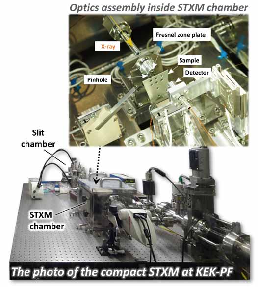
Compact STXM at KEK-PF
The design of STXM is flexible, it can be customized to suit the requirements of the beamline. For instance, in order to avoid low frequency floor vibration, STXM is generally embedded into the top of the polymer-granite, and permanently installed at the end-station of a soft x-ray imagining beamline. However, by adequate modification, it is possible to make the instrument portable. The KEK-PF STXM (see right) has a particular feature of an all-in-one breadboard, which means all the STXM components from the virtual source point (slit chamber) to the detection are installed on a single optical table. This compact design allows it to become a portable instrument while its resolution is comparable with the Rayleigh resolution of the zone plate (~ 40nm resolution with 30nm outermost zone plate). [Ref. 1]
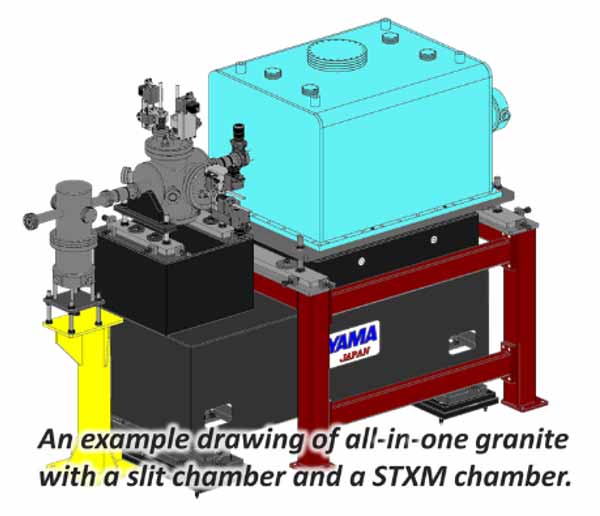
3d model of STXM and slit chamber on granite
TOYAMA is strong in manufacturing and designing various ultra-precision systems for experimentation at worldwide sychrotron radiation facilities. In addition to our experience in STXM and our knowledge in x-ray optics, we are capable to find the ideal solution to customized the STXM for your specific application and turn your idea into reality.
Reference
- Y. Takeichi, N. Inami, H. Suga, C. Miyamoto, T. Ueno, K. Mase, Y. Takahashi, and K. Ono: 'Design and performance of a compact scanning transmission X-ray microscope at the Photon Factory', Rev. Sci. Instrum. 87, 013704 (2016).